Blood and blood circulation
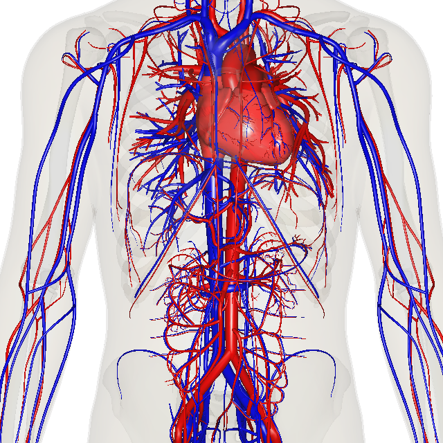
Blood is a vital fluid that transports oxygen, nutrients, and hormones throughout the body. It comprises plasma, red blood cells (carrying oxygen), white blood cells (fighting infections), and platelets (aiding clotting). The circulatory system, powered by the heart, ensures efficient blood flow through arteries (carrying blood away from the heart), veins (carrying blood towards the heart), and capillaries (facilitating exchange of substances). This intricate system is crucial for maintaining homeostasis and overall bodily functions.
Note: This is a very brief overview. For TNPSC exams, you’ll need to study the topic in greater depth. Blood and blood circulation notes taken 10th Samacheer Science book as follows. Also attached 10th and 11th samacheer book pdf at the enad for your reference for the same topic.
Blood
Blood is the main circulatory medium in the human body. It is a red coloured fluid connective tissue.
Components of Blood
The blood consists of two main components. The fluid plasma and the formed elements (blood cells) which are found suspended in the plasma.
Plasma
It is slightly alkaline, containing non-cellular substance which constitutes about 55% of the blood. Organic substances like proteins, glucose, urea, enzymes, hormones, vitamins and minerals are present in the plasma.
Formed Elements of Blood
Blood corpuscles are of three types:
- Red blood corpuscles (RBC) or Erythrocytes
- White blood corpuscles (WBC) or Leucocytes
- Blood platelets or Thrombocytes
Red blood corpuscles (Erythrocytes)
They are the most abundant cells in the human body. RBCs are formed in
the bone marrow. The RBCs impart red colour to the blood due to presence of respiratory pigment haemoglobin.
Matured mammalian RBCs do not have cell organelles and nucleus. They are
biconcave and disc-shaped. Their life span is about 120 days. RBC is involved in the transport of oxygen from lungs to tissues.
Why does mammalian RBC lack cell organelles and nucleus?
Mammalian RBC lack nucleus and makes the cells biconcave and increase surface area for oxygen binding, loss of mitochondria allows the RBC to transport all the oxygen to tissues, and loss of endoplasmic reticulum allows more flexibility for RBC to move through the narrow capillaries.
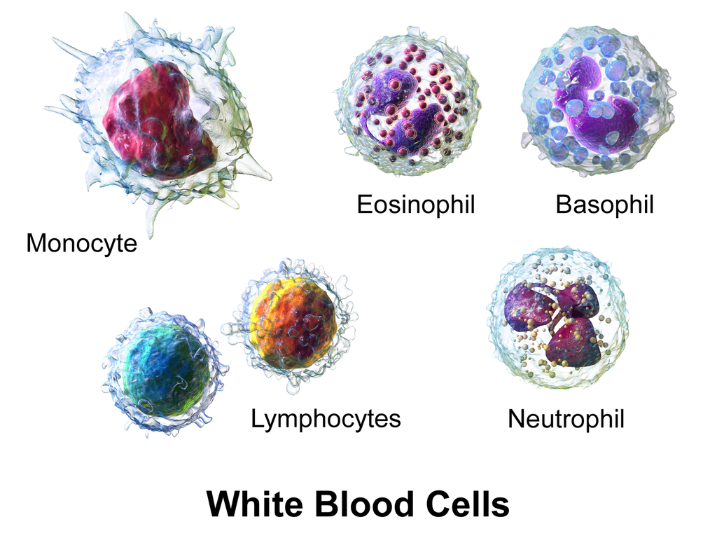
White blood corpuscles (Leucocytes)
WBC’s are colourless. They do not have haemoglobin and are nucleated cells. It is found in the bone marrow, spleen, thymus and lymph nodes. They are capable of amoeboid movement.
The white blood corpuscles can be grouped into two categories:
- Granulocytes
- Agranulocytes
Granulocytes
They contain granules in their cytoplasm. Their nucleus is irregular or lobed. The granulocytes are of three types:
- Neutrophils
- Eosinophils
- Basophils
Neutrophils
They are large in size and have a 2 – 7 lobed nucleus. These corpuscles form 60% – 65% of the total leucocytes. Their numbers are increased during infection and inflammation.
Eosinophils
It has a bilobed nucleus and constitute 2% – 3% of the total leucocytes. Their number increases during conditions of allergy and parasitic infections. It brings about detoxification of toxins.
Basophils
Basophils have lobed nucleus. They form 0.5-1.0% of the total leucocytes. They release chemicals during the process of inflammation.
Agranulocytes
Granules are not found in the cytoplasm of these cells. The agranulocytes are of two types:
- Lymphocytes
- Monocytes
Lymphocytes
These are about 20-25% of the total leucocytes. They produce antibodies during bacterial and viral infections.
Monocytes
They are the largest of the leucocytes and are amoeboid in shape. These cells form 5 – 6 % of the total leucocytes.They are phagocytic and can engulf bacteria.
Blood Platelets or Thrombocytes
These are small and colourless. They do not have nucleus. There are about 2,50,000 – 4,00,000 platelets / cubic mm of blood. Life span of platelets is 8–10 days. They play an important role in clotting of blood. Platelets form clot at the site of injury and prevent blood loss.
More to Know
Anemia: Decrease in number of erythrocytes.
Leucocytosis: Increase in the number of leukocytes.
Leukopenia: Decrease in number of leukocytes.
Thrombocytopenia: Decrease in the number of thrombocytes.
Functions of blood
- Transport of respiratory gases (Oxygen and CO2).
- Transport of digested food materials to the different body cells.
- Transport of hormones.
- Transport of nitrogenous excretory products like ammonia, urea and uric acid.
- It is involved in protection of the body and defense against diseases.
- It acts as buffer and also helps in regulation of pH and body temperature.
- It maintains proper water balance in the body.
Blood Vessels – Arteries and Veins
Blood vessels are a network of branched tubes that transport blood. There are three types of blood vessels namely arteries, veins and capillaries.
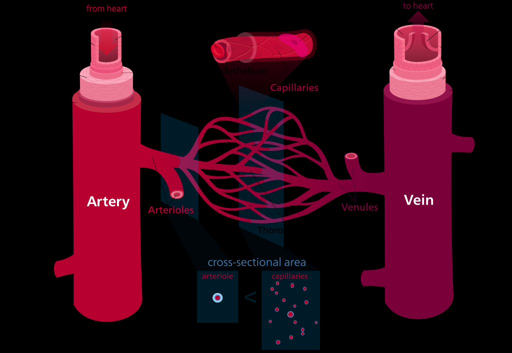
Arteries
They are thick and elastic vessels that carry blood away from the heart to various organs of the body. All arteries carry oxygenated blood except the pulmonary artery which carry deoxygenated blood to the lungs.
Veins
Veins are thin and non-elastic vessels that transport blood to the heart from the different organs. All veins carry de-oxygenated blood except the pulmonary vein which carry oxygenated blood from the lungs to the heart.
Capillaries
Capillaries are narrow tubes formed by branching of arterioles which then
unite to form the venules and veins. They are about 8 µm in diameter. Capillaries are formed of single layer of endothelial cells.
| S.No | Artery | Vein |
| 1 | Distributing vessel | Collecting vessel |
| 2 | Pink in colour | Red in colour |
| 3 | Deep location | Superficial in location |
| 4 | Blood flow with high pressure | Blood flow with low pressure |
| 5 | Wall of artery is strong, thick and elastic | Wall of vein is weak, thin and non-elastic |
| 6 | All arteries carry oxygenated blood except pulmonary arteries | All vein carry de-oxygenate blood except pulmonary veins |
| 7 | Internal valves are absent | Internal valves are present |
Types of Circulatory System
Animals possess two types of circulatory system. They are
- Open type
- Closed type
Open type
In open type the blood is pumped by heart into blood vessels that open into blood spaces called as sinuses. These sinuses are the body cavities which are called haemocoel. Capillary system is absent. e.g. Arthropods, Molluscs
and Ascidians.
Closed type
In closed type the blood flows in a complete circuit around the body through specific blood vessels. The blood flows from arteries to veins through small blood vessels called capillaries. e.g Vertebrates.
More to Know
Closed circulatory system was discovered by William Harvey (1628) who is regarded the Father of Modern Physiology.
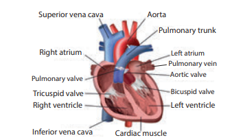
Structure of Human Heart
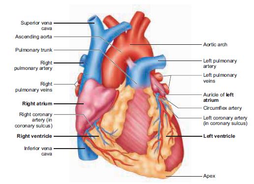
Heart is a muscular pumping organ that pumps out the blood into the blood vessels. Human heart is situated between the lungs, slightly tilted toward the left and above the diaphragm in the thoracic cavity. The heart is made of specialized type of muscle called the cardiac muscle.
The heart is enclosed in a double walled sac called pericardium. It contains lubricating pericardial fluid which reduces friction during heart beat and protects it from mechanical injuries.
The human heart is four chambered. The two upper thin walled chambers of the heart are called auricle or atria (sing: atrium) and two lower thick walled chambers are called ventricles.
The chambers are separated by partition called septum. The septum between
auricles and ventricles prevents the mixing of oxygenated and deoxygenated blood.
The two auricles are separated from each other by interatrial septum.The left atrium is smaller than the right atrium. The right atrium receives deoxygenated blood from different parts of the body through the main veins superior vena cava, inferior vena cava and coronary sinus.
Pulmonary veins bring oxygenated blood to the left atrium from the lungs. The right and left auricles pump blood into the right and left ventricles respectively.
The ventricles form the lower part of the heart. The two ventricles are separated from each other by an interventricular septum. The left and right ventricles have thick walls because the ventricles have to pump out blood with force away from the heart.
From the right ventricle arises the pulmonary trunk which bifurcates to
form right and left pulmonary arteries. The right and left pulmonary arteries supply deoxygenated blood to the lungs of the respective side.
The left ventricle is longer and narrower than the right ventricle. The walls are about three times thicker than the right ventricle. The left ventricle gives
rise to aorta. The oxygenated blood is supplied by the aorta to various organs of the body. The coronary arteries supply blood to the heart.
Valves
The valves are the muscular flaps that regulate the flow of blood in a single
direction and prevent back flow of blood. The heart contains three types of valves.
Right atrioventricular valve
It is located between the right auricle and right ventricle. It has three thin triangular leaf like flaps and therefore called tricuspid valve. The apices of the
flaps are held in position by chordae tendinae arising from the muscular projection of the ventricle wall known as papillary muscles.
Left atrioventricular valve
It is located between the left auricle and left ventricle. It has two cusps and therefore called bicuspid or mitral valve.
Semilunar valves
The major arteries (pulmonary artery and aorta) which leave the heart have semilunar valves which prevent backward flow of blood into the ventricles. They are the pulmonary and aortic semilunar valves.
More to Know
Heart chambers in vertebrate animals
Two chambered: Fishes
Three chambered: Amphibians
Incomplete four chambered: Reptiles
Four chambered: Aves, Mammals and Crocodiles (Reptile)
Types of Blood Circulation
The blood circulates in our body as oxygenated and deoxygenated blood.
The types of circulation are:
Systemic circulation
Circulation of oxygenated blood from the left ventricle of the heart to various organs of the body and return of deoxygenated blood to the right atrium. Aorta carries oxygenated blood to all the organs of the body.
Pulmonary circulation
The path of pulmonary circulation starts in the right ventricle. Pulmonary artery arises from the right ventricle and reaches the lungs with deoxygenated blood. Pulmonary veins collect the oxygenated blood from the lungs and supplies it to the left atrium of the heart.
Coronary circulation
The supply of blood to the heart muscles (cardiac muscles) is called as coronary circulation. Cardiac muscles receive oxygenated blood from coronary
arteries that originate from the aortic arch.
Deoxygenated blood from the cardiac muscles drains into the right atrium by the coronary sinuses.
When the blood circulates twice through the heart in one complete cycle it is called double circulation. In double circulation the oxygenated blood do not mix with the deoxygenated blood.
However, in some animals the oxygenated and deoxygenated blood are mixed and pass through the heart only once. This type of circulation is called single circulation. e.g., fishes, amphibians and certain reptiles.
Conclusion
In conclusion, the intricate interplay between Blood and blood circulation is fundamental for sustaining life. The heart’s relentless pumping propels blood through a vast network of vessels, ensuring the continuous delivery of oxygen and nutrients to every cell while efficiently removing waste products. Understanding the complex mechanisms of blood circulation is crucial for comprehending various physiological processes and their implications for human health.
HI BROTHER, YOUR NOTES ARE VERY HELPUL